


Osteochondrosis is a degenerative-dystrophic spinal disease, basically a damage to the intervertebral disc.The development of spinal degenerative disease is facilitated by prolonged microtraumatization, excessive static and dynamic burden, hereditary tendency, advanced age.The most common localization of the wound is the cervical and lumbar spine.This is due to their biggest mobility and burden.
General concept of osteochondrosis
The intervertebral disc from time to time loses its fluid and loss of its shock function.It becomes less resistant to physical energy.The fibrous ring, located on the edge of the disc, gradually thinner, cracked in it.Pulpic nucleus shifts along the edge of the cracks and shapes formedProtub(Local protrusion, 1 degree).Due to intensive physical activity, highlights can increase and move into the lumen of the vertebral tract.In this case, they talk about disc hernia (2 degrees).Sometimes -nucleus -free fragments can be formed -Sequesters.
In the early stages of the disease, the pain can be explained by overcoming the fibrous ring and the posterior longitudinal ligament irritation.Pain can be localized locally on the back or neck, as well as in remote areas.With cervical osteochondrosis, pain can be reflected in the back of the head, blades and intererspace areas, shoulder and hand carriers.
The pain is accompanied by a spasm of the segment muscle reflex.This phenomenon has a protective properties and stabilizes the part defined from the spinal column.Over time, muscle contraction is a source of free pain.When moving to the intervertebral hole, the hernia squeezes the neighbor's nerve root.Radicular pain has a shooting, absorbed, clearly localized character during nerve preservation.It is accompanied by appropriate neurological manifestations:
- decrease in sensitivity;
- reflex failure;
- muscle weakness.
The disc degeneration violates the normal anatomical ratio between the components of the spinal column: disc, vertebra, joints and ligaments.The gradual decrease in the height of the intervertebral disc leads to changes in articular relationships and the formation of subluxation and vertebral dislocation.This fact shows the instability of the spinal column and reduces the durability of the injury, which can cause the severity of osteochondrosis.
With age, spinal stability is restored due to the formation of osteophytes, articular hypertrophy, disc fibrosis, articular ligaments and capsules.The final stage of the pathological process is called spondylosis.The pain at this time is slowing down.
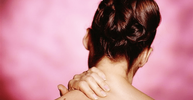
The main symptoms of cervical osteochondrosis
At the level of the cervical segment, their nerve and arterial roots, the spinal cord and its vessels, and the spinal artery can be compressed.Spinal cord compression may be caused by posterior intervertebral hernia or rear osteophytes.People with narrow vertebral channels are very exposed to this.With hernia, the signs of cervical osteochondrosis compression develop quickly, and the symptoms of the cerebrospinal fluvine current block are softer.
It is very difficult to clinically distinguishing the spinal cord compression with tumors and hernia.Cervical spinal osteochondrosis is indicated by spastic paresis of the foot, impairment of sensitivity, pain and weakness in the hands.In some cases, compression signs are combined with signs of spinal cord ischemia arising from compression of the spinal artery and radicular vessels.
Symptoms of damage to anterior horns and departments of the ventral department may arrive -it develops with the involvement of the pyramid path (blood supply to the anterior spinal artery).Anterior spinal syndrome occurs: slow arm paresis, foot spastic paresis, impaired sphincter function.Sometimes the symptoms of rough violations of deep sensitivity in the hands develop.After 2-3 weeks, signs of spinal stroke begin to decline.In terms of pathological focus, we can talk about the severity of the residual phenomenon.
Cervical myelopathy
Myelopathy is a chronic ischemical for cervical osteochondrosis.The major role in the development of this syndrome is played by compression of blood vessels.The most feature is the defeat of the ventral part of the side pole and the front horn.It is indicated by spastikoatrophic paresis from the arm, spastic paresis, deep sensitivity of the foot (classical triad).
In some patients, the symptoms of lermitta appear: the feeling goes beyond electrical release along the spine with the irradiation of pain in the hands and feet on the way.It is possible to develop a side -by -side amyotropic sclerosis where there is no symptoms of bulbar.
An important role in myalopathy confirmation is played by MRI and CT, which reveals compression of shell bags with osteophytes and a bunch of yellow.
Signs -Radicular compression signs
Because the underlying disc is faster, spondylarthrosis develops in the corresponding segment.Osteophytes narrow the intervertebral holes and squeeze the roots (at the lumbar level more often the compression of the disc hernia in the epidural space).When moving the growth head, the spine is injured, which causes the formation of edema, which further narrows the intervertebral hole.Develop reactive inflammatory response.
Clinical manifestations:
- C3 -Koreshok (under 2 cervical vertebrae, occurs relatively) - pain in the same half, swollen tongue, coma in the throat;
- C4 -Koreshok - pain in the proper shoulder flow, klavikun, trapezoid muscle atrophy, decreased neck muscle tone (irritation 3 and 4 cervical roots increases the diaphragm tone, leading to a shift in the liver and the appearance of papardical angina);
- C5 -decor - pain in the neck and outer surface of the shoulder, deltoid muscle hypotrophy;
- C6 -Koreshok (one of the most common localizations) -pain in the neck, blade, shoulder, radial surface spreads to 1 finger, parepezesia in the hands, weakness of the two -head muscle;
- C7-Koreshok-Pain spreads to 2-3 fingers, accompanied by paresthesia, three-head muscle weakness;
- C8 -Koreshok -Pain extends to the surface of the lower arm to the 5th finger, accompanied by paresthesia.
Cervical reflex syndrome
Vertebral syndrome is characterized by acute cervical pain (bastard, cervix), less chronic or subacute pain.The main sources of the pain syndrome are fibrous rings, back longitudinal ligaments, joint capsules, tense muscles.Krivosheya is not very pronounced as a spinal curvature at the lumbar level.
Sickness, radiating to the back of the head.Intensified while driving or prolonged stay in one position.In palpation, the pain of the spinous process and the joint capsule on the sore side (along the posterior surface of the neck 3-4 cm is the side of the spicy process).Involvement in the process not only the back, but also the front muscle of the spine (anterior staircase, etc. -other) is characteristic.
Anterior ladder syndrome
Staircase muscle tension often occurs with cervical osteochondrosis.The muscles are determined by the sternum-coat muscle in the form of pressure grade, compact and increased size compared to a healthy side.Due to voltage, supravic vessel compression occurs, which is accompanied by pain and swelling in the hands, impaired sensitivity and motor activity (along the elbow nerve).Pain is increasing in horizontal position.
Small chest muscle syndrome
The mechanism of development is similar to the previous one.Vascular freezing beam compression occurs between the muscles and the bone (or the corave process) in the event of an increase in the abduction.It is accompanied by pain in the chest, shoulder blades, hands.
Existing features are often considered to be pain in the heart with VSD (no acute attacks, the effects of taking nitroglycerin or sedatives no, increased symptoms during movement and palpation of pain points).
The back sympathetic syndrome
Distropic disorders, vasomotor caused by the vertebral artery plexus plexus irritation are characteristic.The plexus branch is located in the brain and skull tissue.It is clinically indicated by dizziness, ringing in the ear, unusual disorders, anxiety.
Vertebral artery compression with osteophytes derived from the spinal column joint, combined with atherosclerotic damage to the vessels, is an important pathogenetic factor in the development of the brain and spinal cord deficiency.
Conclusion
In most cases, pain in the hands and neck is associated with cervical osteochondrosis.In some patients, the pain is caused by intervertebral disc hernia, in other -other - osteophytes and spinal joint arthrosis.Each of these options can cause local pain or reflected, radicular syndrome and myelopathy.When examining the patient with pain in the neck, it is necessary to exclude pathology such as:
- spinal tumor;
- epidural abscess;
- spondylitis;
- subarachnoid bleeding;
- Meningitis;
- Hall abscess;
- carotid artery stratification;
- Cervical spine fracture.